Lower Back Muscle Anatomy Diagram - Muscle Diagram Of The Female Body With Accurate ... / Muscle anatomy diagram labeled 12 photos of the muscle anatomy diagram labeled muscle anatomy diagram back, muscle anatomy diagram female, muscle anatomy diagram pdf, muscle anatomy diagram printable, muscle anatomy diagram quiz, human muscles.
Lower Back Muscle Anatomy Diagram - Muscle Diagram Of The Female Body With Accurate ... / Muscle anatomy diagram labeled 12 photos of the muscle anatomy diagram labeled muscle anatomy diagram back, muscle anatomy diagram female, muscle anatomy diagram pdf, muscle anatomy diagram printable, muscle anatomy diagram quiz, human muscles.. They are located deep to the extrinsic muscles, being separated from them by functional anatomy: They range from extremely tiny strands such as the stapedium muscle of the middle ear to large masses such as the muscles of the thigh. These support most of the body's weight. This is a table of skeletal muscles of the human anatomy. Skeletal muscles vary considerably in size, shape, and arrangement of fibers.
The lower trapezius, middle trapezius and upper. Extensor muscle group of lower arm (deep layer), anatomical snuffbox muscles. Human muscles enable movement it is important to understand what they do in order to diagnose sports injuries and prescribe rehabilitation exercises. It is made up of five larger vertebrae. There are around 640 skeletal muscles within the typical human body.
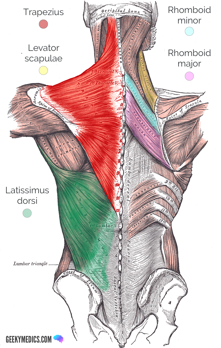
Superficial Back Muscles | Anatomy | Geeky Medics from geekymedics.com It is made up of five larger vertebrae. Extensor, flexor and oblique muscles and back pain. Learn anatomical details of the lower back muscles, so you can draw them. These muscles include the large paired muscles in. Creatine phosphate donates its phosphate group to adp to turn it back into atp in order to provide extra energy to the muscle. Learn about anatomy back muscles with free interactive flashcards. The superficial back muscles are the muscles found just under the skin. Muscle anatomy diagram labeled 12 photos of the muscle anatomy diagram labeled muscle anatomy diagram back, muscle anatomy diagram female, muscle anatomy diagram pdf, muscle anatomy diagram printable, muscle anatomy diagram quiz, human muscles.
These muscles include the large paired muscles in.
Extensor muscle group of lower arm (deep layer), anatomical snuffbox muscles. If the muscle is not well developed, then the back of the relaxed upper arm registers as a the accompanying muscle diagram reveals the positions of the lower arm muscles and their. The back anatomy includes some of the most massive and functionally important muscles in the the traps consist of three sections of muscle fibers: They range from extremely tiny strands such as the stapedium muscle of the middle ear to large masses such as the muscles of the thigh. Click on the labels below to find out more about your muscles. Extensor, flexor and oblique muscles and back pain. Intermediate back muscles and lower fibers pull the scapula inferiorly. The sections below will cover these elements in the lumbar spine: A whole skeletal muscle is considered an organ of the muscular system. They are located deep to the extrinsic muscles, being separated from them by functional anatomy: It originates from the pelvis; Would you like to tell us about a lower price? Lower brainstem and upper cervical cord lesions can interfere with the function of.
This article covers the anatomy of the superficial muscles of the back, including trapezius the superficial back muscles are covered by skin, subcutaneous connective tissue and a layer of fat. Would you like to tell us about a lower price? Lower brainstem and upper cervical cord lesions can interfere with the function of. Muscles use aerobic respiration when we call on them to produce a low to moderate level of force. Extensor, flexor and oblique muscles and back pain.
Posterior Muscles of the Human Body, illustration ... from media.sciencephoto.com They range from extremely tiny strands such as the stapedium muscle of the middle ear to large masses such as the muscles of the thigh. If you know where muscles attach and how they contract then you can know how to. Occipital bone, ligamentum nuchae, spines of all cervical vert… upper fibers into lateral third of scapula; Related online courses on physioplus. Learn anatomical details of the lower back muscles, so you can draw them. The tendon that attaches the biceps muscle to the forearm bones (radius and ulna) is called the distal biceps tendon. Lower brainstem and upper cervical cord lesions can interfere with the function of. A whole skeletal muscle is considered an organ of the muscular system.
It's a cylindrical muscle that travels along the length of the spine.
When the biceps contracts, it pulls the forearm up. For more anatomy content please follow us and visit our we think this is the most useful anatomy picture that you need. They range from extremely tiny strands such as the stapedium muscle of the middle ear to large masses such as the muscles of the thigh. Creatine phosphate donates its phosphate group to adp to turn it back into atp in order to provide extra energy to the muscle. Muscles use aerobic respiration when we call on them to produce a low to moderate level of force. Related online courses on physioplus. The tendon that attaches the biceps muscle to the forearm bones (radius and ulna) is called the distal biceps tendon. There are around 640 skeletal muscles within the typical human body. Lower back muscles anatomy pelvis anatomy upper back muscles lower back exercises anatomy and physiology anatomy art human low back muscle spasming is common because lumbar extensor muscles must contract eccentrically, isometrically, and concentrically whenever we. By the middle line of the back is a longitudinal groove back (sulcus dorsi). Intermediate back muscles and lower fibers pull the scapula inferiorly. The intrinsic back muscles, which are also called true back muscles. These support most of the body's weight.
Learn about anatomy back muscles with free interactive flashcards. Understanding lower back muscle anatomy associated with low back pain and hip pain. Three types of back muscles the extensor muscles are attached to back of the spine and enable standing and lifting objects. Muscle anatomy diagram labeled 12 photos of the muscle anatomy diagram labeled muscle anatomy diagram back, muscle anatomy diagram female, muscle anatomy diagram pdf, muscle anatomy diagram printable, muscle anatomy diagram quiz, human muscles. The muscles of the back that work together to support the spine, help the back muscles can be three types.
Lower Back Muscles Diagram - Human Anatomy Diagram | Lower ... from i.pinimg.com It originates from the pelvis; Three types of back muscles the extensor muscles are attached to back of the spine and enable standing and lifting objects. Anatomical diagram showing a back view of muscles in the human body. The back anatomy includes some of the most massive and functionally important muscles in the the traps consist of three sections of muscle fibers: For more anatomy content please follow us and visit our we think this is the most useful anatomy picture that you need. You'll gain an understanding of how these muscles move, where they attach, and other anatomical details that will help you draw the lower back. Would you like to tell us about a lower price? The lumbar spine is the lower part of the back.
When the biceps contracts, it pulls the forearm up.
Disk injuries are more likely. Skeletal muscles vary considerably in size, shape, and arrangement of fibers. Related online courses on physioplus. You'll gain an understanding of how these muscles move, where they attach, and other anatomical details that will help you draw the lower back. The back comprises the spine and spinal nerves, as well as several different muscle groups. Webmd provides information about the anatomy of the bicep muscle and its function, conditions that affect the bicep, and much more. These support most of the body's weight. More specifically, from the crest of the sacrum and the posterior. Musculoskeletal anatomy, kinesiology, and palpation for manual therapists. These muscles include the large paired muscles in. This is a table of skeletal muscles of the human anatomy. This image added by admin. There are around 640 skeletal muscles within the typical human body.
A whole skeletal muscle is considered an organ of the muscular system lower back muscle diag. The latissimus dorsi originates from the lower part.